Patients first, always.
Examples of saving teeth:
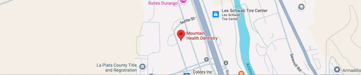
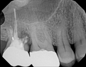
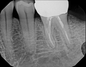
Non-surgical root canal treatment:
 Before
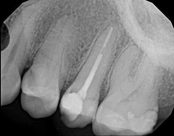 After
After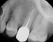 6-month follow-up
The patient presented with mild pain, a cavity, and a broken filling was evident. After performing diagnostic testing, the diagnosis was: irreversible pulpitis, symptomatic periradicular periodontitis. Root canal therapy was done and the tooth healed well and is still functioning.
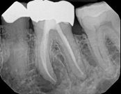
Non-surgical root canal treatment:
 Before
 Immediately After
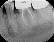 6-month follow-up
This patient presented with pain when chewing. Diagnostic testing was done and determined it was the lower molar. The diagnosis was: pulpal necrosis, symptomatic periradicular periodontitis. Root canal treatment was performed with laser disinfection. The tooth had a crown and we accessed the tooth through the crown. We saved the tooth and the crown.
Apicoectomies, including molar apicoectomies:
 Before
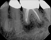 After
After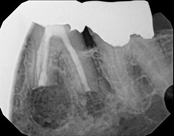 3-month follow-up
This person saw a specialist and was told the molar tooth needed to be extracted because the root canal failed and was infected. The root canal had been done by an endodontic specialist. The diagnosis was: previous endodontic treatment, chronic periradicular abscess. We did an apicoectomy and saved the patient’s tooth.
Mid-root crack repair:
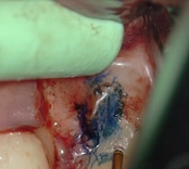 Dye used to verify the crack
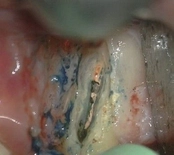 Crack removed
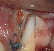 Filled with Geristore
The patient was eating and bit down, felt and heard a “pop.” The tooth had cracked in the middle of the root. The tooth helps hold the patients partial in place and did not want to lose the tooth. The crack was removed with the laser and then restored.

The photograph is 2 months after the crack repair. The area looks like nothing was done (no redness, no scarring, etc.) and the tooth is there helping to hold the partial denture.
Root amputation:
 Before
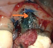 The crack
 After
AfterThis patient had root canal treatment many, many years ago. One root developed a crack all the way through the root leading to an abscess. That root was removed and the tooth is still functionable.
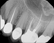
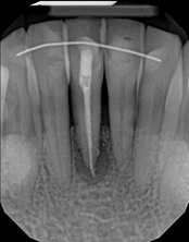
Missed canals and sclerotic orifices/canals:
 Before
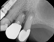 4-month post-op
In this situation, some would say the canals are so “calcified” that root canal treatment can’t be done; or sometimes the canal can’t be found. And because of that the tooth would need an extraction and an implant. In some instances, root canal therapy can’t be done but that does not mean the tooth is a loss. The solution is micro-apical surgery, an apicoectomy.
Root canal retreatment, apicoectomy, and root amputation:
This person had root canal therapy many years ago. One root had excess material outside of the root preventing healing. Another root had a perforation (a hole) and a crack. The large root had a post and had not healed from the initial root canal treatment.
The post was removed, that root was retreated. The root with the excess material, an apicoectomy was done, the excess material, and infection cleaned out. The root with the perforation and crack was removed. The tooth healed and is still in function. This area is a poor area for an implant.
Apicoectomy:
 Before, the failed root canal
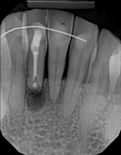 Immediately after the microsuregery
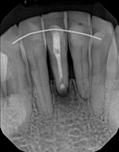 3-months after shows complete healing
The patient was told by a specialist that the root canal failed, the only option was extraction and an implant. She did not want to do that; she wanted to keep her tooth, after all, it is her front tooth. She found us. We performed an apical microsurgery with the laser. The tooth was saved and is still functioning. This is a poor area for an implant, so much better to save the tooth.
Lower molar failed root canal, microsurgery saved the tooth:
 Before
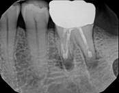 Immediately after
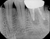 3-month follow-up with complete healing
This patient presented with a failed root canal. The root canal became infected. An apicoectomy (apical microsurgery) was performed using the laser. Patient had minimal swelling, minimal discomfort, and fast healing. The tooth was saved and an implant avoided. This tooth tends to have a lower success rate with root canal therapy; thankfully apical microsurgery was available to save the tooth.
At the 3 month follow-up, the gums look like nothing was done thanks to the skill and use of the laser for the procedure.
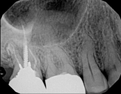
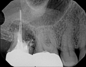
Upper molar failed root canal and apicoectomy:
 Before
 Immediately after
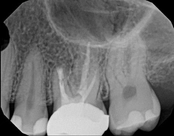 3-month follow-up
This person presented with one root that had failed from root canal treatment. The arrow in the picture, shows the infection as well as extra filling material that was pushed out of the root. An apical microsurgery was performed using the laser. The infection and excess material was cleaned out. Complete healing is visible 3 months after. The tooth was saved. This is a poor area for an implant.